The weird and wonderful: A case of a calf born with extra legs
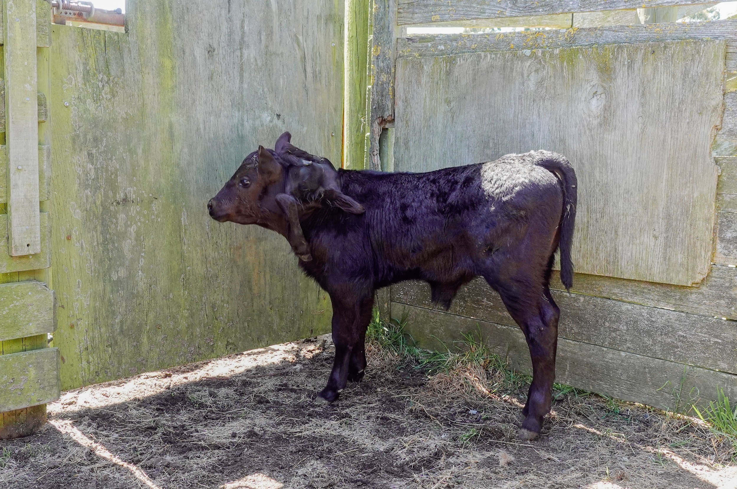
Meet Spider (the name given to him by the RVC vets), an Angus calf born with 3 extra legs!

WATCH: Spider’s CT Scan, surgery and recovery!
Dr John Spearpoint, one of RVC’s large animal vets, was called by a farmer when he noticed a calf with an usual lump on the back of his neck. The calf was walking around normally and suckling from his mother. On examination, three different sized limbs were hanging from the top of the neck attached to a large firm mass. The limbs were smaller than normal limbs and only partially formed, characterized by a single toe and joints arranged at unusual angles. There was no movement or nerve sensation in the accessory limbs. As a salvage procedure, the farmer was interested to know if the mass could be removed.
Spider the calf with his mass and accessory limbs.
There was no movement or nerve sensation in the accessory limbs.
The calf was brought into the clinic for further investigation. Spider was sedated and placed in the CT machine to determine how the accessory limbs were attached to the neck and if surgery could be safely undertaken given close proximity to the spinal vertebrae.
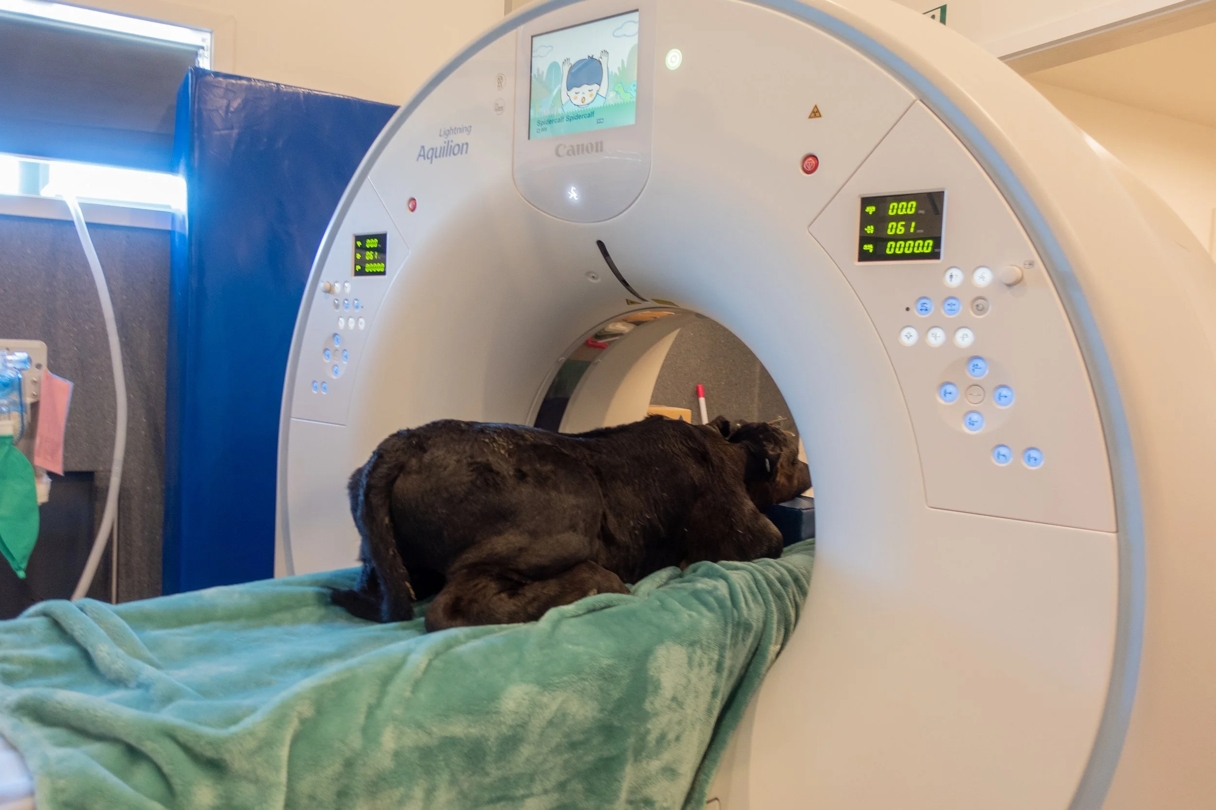
Spider, sedated, in the CT Scanner.

The CT Scan in action. Vet Bryn assisted with CT imaging diagnostics.

Cross-sectional image of the mass.

Soft tissue CT image of the mass.

3D view of Spider’s skeleton, including the bones in the accessory limbs.

Spider was placed on an anaesthetic machine and surgical amputation of the limbs and mass were performed. The mass consisted of a large amount of fibrous tissue and the accessory limbs were inter-twined with no attachment to the underlying skeleton. After a long surgery, Spider recovered smoothly.
Later that day Spider returned to the farm and his mother. Sporting a large scar, he happily returned to grazing in the paddock. Post-operative care included ongoing pain relief and antibiotic injections for a week.
Several months later, Spider’s progress was unbelievable. He continued to graze normally and put on weight. Here he is pictured with the pink ear tag – it is hard to tell the difference from other calves born around the same time.


So, what likely caused this?
Congenital abnormalities like this occur during foetal development of the musculoskeletal system. Polymelia is the term given to the development of extra limbs, which is further classified according to the region of the body where accessory limbs are attached. Notomelia correctly describes Spider’s condition, where the extra limbs were attached in the region of the embryonic notochord (an area that later develops into the vertebral column).
These conditions occur very early in the developing embryo and may be due to a heritable genetic mutation or from environmental influences such as exposure to toxins, chemicals or infectious agents like viruses during pregnancy. In Spider’s case, given the Angus breed predisposition and later DNA analysis, we confirmed he had a genetic condition called “Development Duplication (DD)”.
DD is an inherited recessive genetic condition where both parents are carriers of a defective recessive gene. This is a recognised recessive condition in the Angus breed, and although not all Angus cattle are carriers, it has been traced back to a popular sire used heavily in stud breeding programs due to his desirable carcase traits.
In humans, recessive genetic conditions like colour blindness and cystic fibrosis are similar examples that can occur in offspring when both normal parents carry recessive genes.
Is this likely to happen again?

Free – animal is normal and carries two copies of the normal form and no copies of the mutation.
Carrier – animal which looks normal, but carries one copy of the mutation which can be passed onto offspring.
Affected – affected or abnormal animal which carries two copies of the mutation.
Polymelia is an extreme manifestation of this recessive genetic condition and several cases have been reported in literature. Many cases may go undetected as early embryonic loss is possible.
Genetic studies into this condition indicate that the DD mutation gene is transmitted with incomplete penetrance meaning not all calves born from carrier parents will show signs of this condition and will appear as normal animals. However, using the principles of Mendelian genetic inheritance which determines how traits are passed from parents to offspring, every time you breed a carrier to a carrier, there is a 25% chance the calf may or may not express this condition. There is also a 50% risk the calf will carry the DD mutation but otherwise appear normal; and a 25% chance the calf is a non-carrier and appears normal.
Registered Angus studs now screen animals for known recessive genetic conditions as part of the registration of their animals with the Angus breed society to limit further spread of these conditions. If you’re intending on buying an Angus bull, ask the vendor if a DNA check for recessive genetic conditions has been undertaken or has been assessed as being a non-carrier through parentage evaluation. Only animals with an ancestry linkage to a known carrier are at risk.