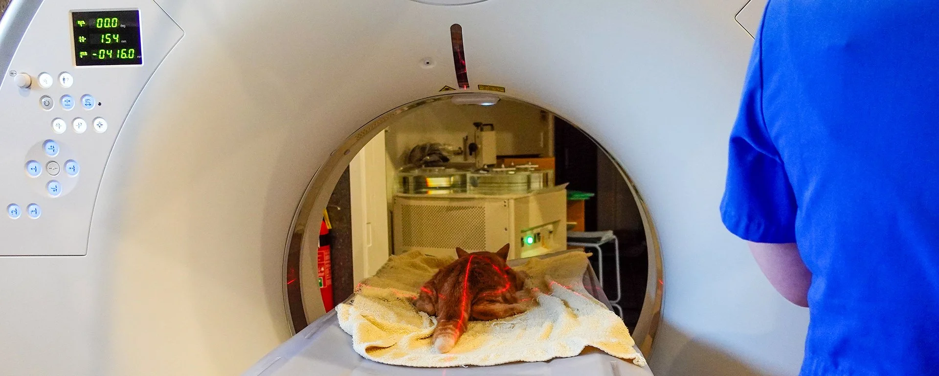
CT Scanning at RVC

WATCH: Hear from Vet Bryn and Vet Nurse Nicole, as they talk about our CT scanner, how it works, and how it will improve diagnostic and pre-surgical planning for pets in the Canterbury region.
We are proud of our NZVA Best Practice and Hospital Standard Accreditation, along with our ISFM Gold Standard Cat Friendly Status. We are committed to a high standard of care for clients and their pets.
We strive for Fear Free Handling with our patients to ensure a stress free visit. Our patient focused team ensure our post operative patients are pain free and have a comfortable recovery.
We have dedicated emergency and critical care vets and nurses 24/7 to provide intensive care to compromised patients.


Our Aquilion Lightning CT scanner has the industry's widest gantry to accommodate companion animals through to equine.

We offer contrast studies and 3D views to help improve pre-surgical planning.
Pet Owner FAQs
-
CT is a specialised form of X-ray scanning enabling cross-sectional images of your pet to be produced, providing more detailed information for your vet. A special contrast agent (dye) is sometimes used which may be injected into your pet’s bloodstream or around other areas of interest to highlight increased blood flow or changes to their anatomy. CT is performed under general anaesthesia or heavy sedation.
-
There are many reasons why your pet might need a CT examination. CT is commonly used to detect cancerous growths and determine the extent of any spread. It is also helpful for assessing orthopaedic issues, including elbow dysplasia in young large-breed dogs. It can also be used to better understand:
· complex fractures (including of the pelvis)
· identify the cause of nasal discharge
· investigate persistent ear infections and coughs
· if foreign material is lodged somewhere, causing discharge or swelling
-
Alternatives to CT include Magnetic Resonance Imaging (MRI) and standard radiography (X-rays), the latter remaining the mainstay of most investigations into illness and injury.
X-rays are a quick and relatively straightforward procedure providing useful information. However, X-rays usually provide less information than CT and are often difficult to interpret as structures appear layered over one other.
As CT can provide us with thin slice images, you can understand how this provides much more specific, clearer information.
MRI can be more useful than CT in some cases because it allows better visualization of soft tissue structures in certain areas (such as the brain and spinal cord). However, MRI is less readily available, more expensive and takes longer to acquire the images (sometimes up to 30 mins). CT images can be produced within seconds to minutes, which is one reason why CT is more useful for imaging the chest as movement from breathing often blurs MRI images.
-
People who perform CT examinations abide by strict legal regulations, limiting their exposure to X-rays which are a form of radiation. X-rays can potentially cause genetic effects (mutations) in the body that can lead to cancer.
Your pet will not be exposed to enough X-rays during the CT procedure to incur risk of radiation damage either immediately or later in life. The regulations mean that both staff and owners are not allowed to hold patients for CT examinations or be in the room whilst the images are being obtained. This means your pet will need to be anaesthetised or heavily sedated for the procedure.
Anaesthetics and sedatives do carry some risk, and the team will discuss these with you. Risks must always be weighed against potential benefits gained by performing the examination. The contrast agent used for these examinations is excreted from the body in the urine and may not be appropriate in animals with severe renal disease. Your vet or nurse will also discuss this with you. Although extremely rare, an allergic reaction to the contrast agent can occur.
-
Eating and drinking: as your pet will be anaesthetised or sedated to perform the CT examination, you will need to withhold food on the day of the examination to reduce the chances of reflux or involuntary vomiting. Water may be given until you arrive at the practice.
Toileting: please take your dog out before your visit to the clinic and encourage them to empty their bladder and bowel. Cats should be provided with a litter tray until you leave for your appointment.
Medication: if your pet is on medication, you should check with your vet whether it should be given on the morning of the procedure. Medicines such as insulin may need to have an adjusted dose prior to admission.
-
You do not need to bring anything other than your pet! A small amount of a familiar food can be useful to tempt them to eat after the procedure.
-
A member of the hospital team will admit your pet, go over the consent form confirming that you understand the procedure and risks involved. They will discuss if any further treatment or tests are required and ask if you have any questions.
It is important you provide contact details, ensuring someone is available to answer a call from the practice in case urgent information is required.
-
Your pet is likely to stay with us for the day and will be discharged in the late afternoon or early evening. This allows time for recovery from the anaesthetic or sedative.
If surgery is to be performed at RVC on the same day, your pet will often need to stay overnight. Our clinic is staffed 24 hours a day allowing us to take good care of patients around the clock.
-
Your pet will be anaesthetised or heavily sedated and will be monitored throughout the procedure by a vet and a trained veterinary nurse.
Monitoring is performed remotely during the scan as no one is allowed in the examination room whilst the CT machine is operational.
During the CT procedure, your pet will pass through the scanner and X-rays taken from multiple angles are obtained. This information is processed by a powerful computer to create the images.
The process starts with a very quick, simple X-ray of the patient (topogram) allowing us to hone in on the area of interest. After the initial images, a contrast agent is often injected into a small catheter placed in one of your pet’s limbs and the process repeated.
After the examination your pet will be allowed to recover from the sedative or anaesthetic in a comfortable cage and is monitored until fully awake when they are offered food and water.
The CT images will sometimes be interpreted by your vet but are usually sent to a radiologist for their opinion. The referral process may mean that results are not available for a few days. Once the report has been received your vet will contact you to discuss the findings. Contact your vet clinic if you have not heard from them within a week so that they can investigate any delay.
-
Your pet will require some care and attention once they are discharged, our team will discuss these needs specifically.
They will have been offered a small meal after recovery but are more likely to eat familiar foods in their home environment, so they should be offered food and water as soon as they are settled at home.
Cats should be kept in the house for at least 24 hours (so remember to lock the cat flap).
Dogs should be confined to the garden for a day and then taken out only on the lead for another day.

Interesting CT Scanner case: a calf born with extra legs.
Learn more about Spider’s story - an Angus calf born with three extra legs!