
Root Canal Therapy & Vital Pulpotomy:
Endodontic Therapies an Alternative to Tooth Extraction at RVC
The Dentistry Team at RVC is proud to be able to offer Endodontic treatment as an alternative to tooth extraction for your dog or cat.
Endodontics is a specialized branch of human and veterinary dentistry involving treatment of the nerve and blood supply within the tooth, called the pulp, to preserve tooth function. These procedures serve as a viable alternative to tooth extraction for our pets, particularly for teeth considered structurally or functionally important.
The mandibular (lower) canines in dogs and cats are the most common teeth to receive endodontic therapy. These teeth provide a significant amount of structural support to the lower jaw and the most common complication when extracting these teeth can be accidental jaw fracture particularly in older patients. Other teeth that we encourage saving with endodontics are the maxillary (upper) canines and the main chewing teeth known as the Carnassials of which there are two in the top jaw and two in the bottom jaw.
X-rays under general anesthesia are an essential part of the initial work up to determine if a tooth can receive endodontic therapy. If your pet breaks a bone, it is essential to X-ray the bone to determine the best treatment option. The same goes for a damaged tooth. X-ray findings such as root fractures, abnormal root or pulp anatomy, or advanced periodontal disease would mean extraction is a more appropriate option.
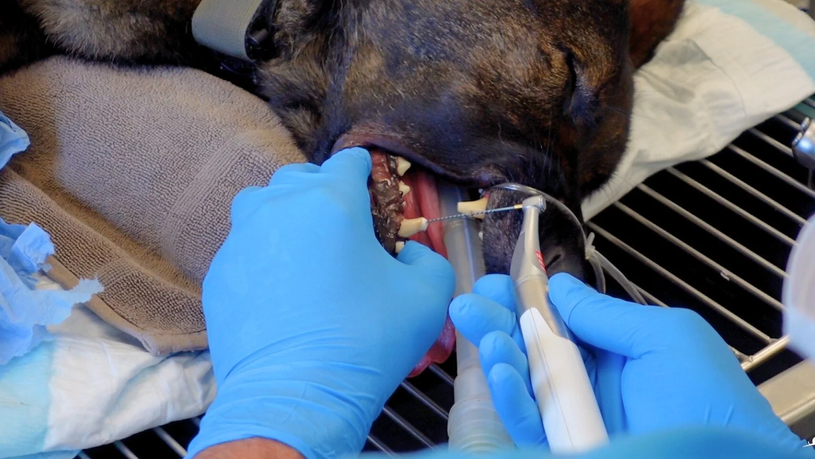
WATCH: Have you ever wondered what happens when a pet receives root canal therapy? Watch Sarge’s procedure to find out more!
What is a Root Canal?
A Root Canal is a procedure recommended for mature teeth that have lost their blood and nerve supply but are otherwise still functional. The root canal procedure involves using specialized instrumentation to remove the diseased pulp, thoroughly clean the canal, fill it with an inert substance called gutta percha then seal the canal to prevent infection. The procedure is far less invasive than extraction with a faster recovery period.
Despite the tooth no longer having blood or nerve supply, it still remains functional allowing the animal to keep the tooth, in most cases, for the remainder of its life. Follow up X-rays once a year for at least 3 years is recommended to monitor the tooth.

A German Shepherd with damaged right lower canine. Note the purple discolouration which indicates pulp damage.

An X-ray of the completed root canal. Red arrow indicates the gutta percha filling in the pulp canal that is bright white on X-ray.

The finished root canal. Red arrow showing the circular hole made to access the pulp canal now sealed over with restorative material to prevent infection.
What is a Vital Pulpotomy?
A Vital Pulpotomy is a procedure recommended for either mature or immature permanent teeth where the intention is to keep the tooth ‘vital’ aka alive. For this procedure the live exposed pulp is ‘patched up’ with a specific material that stimulates the formation of a protective dentinal bridge, keeping the blood and nerve supply intact.
Two reasons we might choose this procedure are due an acute fracture with pulp exposure or as part of a procedure to relieve pain from malocclusion. For an acute fracture, this procedure would need to be done urgently as in within 48 hours of the damage whereas with immature permanent teeth we have up to two weeks in which to perform the procedure. The longer we wait, the less likely this procedure will be successful thus the alternative may be root canal.

A common malocclusion in dogs where the lower canine digs into the palate causing pain when the mouth is closed. The lower canine’s crown height was reduced and at vital pulpotomy performed.

An X-ray of the tooth after Vital Pulpotomy performed. Red arrow indicates material placed to encourage protective dentinal bridge formation.

The finished product showing the crown with a smooth cap covering the pulp for further protection.
Why might we recommend endodontic therapy for your pet?
The most common reason an endodontic therapy may be recommended is due to tooth fracture or pulp damage. Dogs and cats surprisingly have a thinner protective layer of tooth enamel ranging anywhere from 0.1 to 1mm thickness compared to humans which have enamel upwards of 2.5mm! Because of this, dog and cat teeth can be easily fractured by trauma or inappropriate chewing activity. It is important to note that any damage to your pets’ teeth warrants investigation rather than the “wait and see” approach as tooth damage is just as painful in our pets as it is for us.
One other fairly common reason for endodontic therapy, as pictured above, is to relieve a painful malocclusion. Malocclusion means your dog or cat’s teeth aren’t pointing in the right direction so that when they close their mouth, one or more teeth cause damage to other teeth or soft tissues. This can be developmental or due to trauma.
The lower canines tend to be common culprits for this with a condition called “base narrow” canines. The bottom canines grow slightly inwards towards the tongue so that when the mouth closes, the canines dig into the palate creating deep holes, sometimes even going all the way into the nasal cavity (ouch!). An option for correcting this is to reduce the length of the canines and perform a vital pulpotomy. This stops the crown tips from digging into the palate but also keeps their long roots in place to continue to provide bony support.
Any tooth in your pet’s mouth can technically receive endodontic therapy however it is important to note that not every pet is the best candidate. An initial consultation with our veterinary dentist is the most important step as it allows for a detailed discussion around treatment options, potential risks & complications, post operative management, follow up requirements as well as a discussion about cost and payment options.